Hello and welcome to the NVIDIA AI Podcast.
I'm your host, Noah Kravitz. 8 million Americans, and many more globally, visit the emergency room every year complaining of chest pain.
80% of people who undergo a cardiac CT scan The first line test for chest pain show no signs of coronary artery disease.
Yet despite being treated as low risk, New studies show these people die at twice the rate of those with positive scans.
The culprit? Coronary inflammation, which CT scans can't currently spot.
That's where Caristo Diagnostics comes in.
As detailed at the American Heart Association Conference this past November, Caristo says its AI-powered solution is the first technology of its kind able to spot coronary inflammation in cardiac CT scans. currently in use in five NHS hospitals in the UK.
A study of 40,000 patients with chest pain who were followed over 10 years showed Karista's AI accurately revealed coronary inflammation and predicted heart attacks years in advance.
Fully 45% of the patients identified by Caristo's technology had their treatment plan adjusted by physicians.
As someone with a history of heart disease in my own family, I have to say these stats caught my eye and Carrizo's solution sounds pretty incredible.
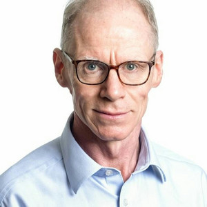
With us today to explain how and why Carrizo's technology works the way it does is Professor Keith Channon.
Dr. Channon is co-founder and chief medical officer at Carissa Diagnostics.
And he is also the chair of cardiovascular medicine at the University of Oxford.
Dr. Channon, thanks so much for joining the NVIDIA AI podcast and welcome.
Thank you so much for the opportunity to talk to you today.
So let's start the conversation off by talking about the clinical problem that Caristo, and I would imagine your work previous to Caristo being founded, is aiming to solve?
Yeah. Well, this is a really big problem because the advent of cardiac CT scans has really helped us understand and detect coronary artery disease, which is the problem that leads to angina and ultimately heart attacks.
And of course, heart attacks even with modern treatments, all too frequently lead to death.
And even if patients survive the heart attack, they may be left with heart damage leading to heart failure and other problems in the long term.
So despite the advances that the cardiovascular community and medical care has made, heart attack risk remains a major global problem.
And it's not just in Western societies, it's also in many of the large emerging economies around the world, especially countries where unfortunately smoking remains very common.
So detecting coronary artery disease has been transformed by the ability to see the coronary arteries on cardiac CT scan.
But the problem that we as clinicians and research scientists based at the University of Oxford started to grapple with is that the narrowing of the coronary artery, the so-called plaque is the long-term consequence of the disease process.
It's not really visualizing the disease itself.
And as you've already mentioned, the biological process that drives the formation of plaque formation in the arteries is inflammation.
And that is caused by elevated cholesterol and diabetes and smoking, all of the so-called risk factors that we've recognized for many years.
And so although the wider use of cardiac CT scans has transformed the diagnostic pathway, Many patients who have a CT coronary scan do not have significant narrowing of their arteries.
They may have minimal plaque formation. They may have no visible plaque formation at all.
And only about 20%, that's one fifth of patients, have arteries which are significantly narrowed.
Those patients, of course, have a very well-defined clinical pathway.
They're seen... They have further investigations and they're treated appropriately.
But for the overwhelming majority of patients... those patients should be assessed for their future risk and treated accordingly.
But what we've found is that inflammation the driver of the disease process is not detected by current blood tests sufficiently precisely to identify individual patients.
And so some of our most recent research work highlights in 40,000 patients who've been followed up for 10 years after having had a CT scan in the past, that although having significantly narrowed coronary arteries is indeed worse than having no significant narrowing in terms of your risk of heart attack,
Because there are fewer patients, most heart attacks and deaths occur in the people who did not have cancer. significantly narrowed arteries at the time of their CT scan.
So what we're doing with CT scans right now is we're detecting a minority of the people who in the next 10 years will have a heart attack or die of a heart attack.
And that's a very sobering thought given... technological advance that we recognize in our CT scanning.
Absolutely. You mentioned the major risk factors, some of the major risk factors that go into assessing someone's risk of, when we talk about heart disease and having a heart attack, is that interchangeable or are they, just to clarify, are they two separate things?
Right, so heart disease is a catch-all term.
Having a heart attack is a very specific event.
A heart attack occurs usually very suddenly, usually abruptly, when a narrowed coronary artery or a coronary artery with inflammation blocks off usually due to the formation of a thrombus or blood clot.
Okay. And that's why a heart attack happens so quickly, so abruptly.
Right. And in fact, this is one of the key points that many people, in fact, more than a half of people who have a heart attack Immediately prior to the heart attack, even though the artery blocked off, the artery was not significantly narrow.
Right. before the heart attack happened.
And that's because inflammation is sufficient to cause the artery to become unhealthy. and be at risk of blocking off very suddenly without warning, even if there's no visible thickening or narrowing of the artery wall,
Cardiology, cardiologists like me and my colleagues were very focused on detecting narrowed or blocked arteries.
And those, to be sure, are very important.
They are not the sole predictor of risk of heart attack or death. conundrum.
That's been the paradox and the challenge for cardiology to overcome.
We've known from biological studies that inflammation is the driver of the disease over years.
It's also... the trigger that leads to the heart attack over minutes or seconds.
And yet we've had no reliable way of detecting inflammation.
We can only see the narrowings or the blockages physically.
How did you go about, you and your colleagues at Oxford, I believe, right, where the research started?
How did you go about looking for a way to... Did you know that the inflammation was the culprit even back then, but you...
Didn't know how to see it? Or how did you narrow it?
What was the problem you narrowed it on?
Right. So the fact that inflammation is a causal factor in the development of coronary artery disease and the precipitation of a heart attack event has been well established by researchers in many laboratories around the world.
Okay. Our breakthrough in the research laboratories at the University of Oxford were to begin to investigate how inflammation in the coronary artery wall affects the fat tissue which surrounds the artery.
And at the time, that wasn't a very scientifically fashionable thing to do.
The focus was on the wall of the artery. and the fat tissue was largely ignored.
What we discovered is that if the coronary artery wall is inflamed, then the surrounding fat tissue undergoes cellular and molecular changes that respond to the inflammation.
And we were able to do that by taking tissue biopsies from patients undergoing open heart surgery.
And in our discovery science research, we analysed molecular signals in the fat tissue to look for signs of inflammation and we found that those signals were readily detectable using molecular techniques, looking at gene expression profiling.
And we also discovered that in the fat tissue, those changes correlated very highly with markers of inflammation and disease in the wall of the coronary artery.
So that was the first breakthrough. When was that?
That was in 2016, 2017. Okay. The next breakthrough was to ask ourselves the question, how could inflammation at the molecular level in the fat tissue surrounding the coronary arteries in people, how could that inflammation in the fat tissue be detected in a way that was clinically useful?
And that's where we turn to cardiac or coronary CT scans, where of course, in order to visualize the coronary arteries, in beautiful detail, the usual practice is to modify the images so that the fat tissue is completely removed and you can see the arteries.
Interesting, right. So for many years, the fat tissue that is present in the imaging data has been excluded and not looked at.
So the breakthrough in our research was to use AI techniques to find associations between invisible radiomic features in the CT scan data and to correlate those with the molecular readouts of inflammation in tissue.
And there are two really big advances here.
One is that the fat tissue around the arteries is very easy to image using CT scanning compared to the very thin wall of the artery.
And secondly, it turns out that those changes that are caused by inflammation in the wall of the artery lead to very striking alterations in the imaging characteristics of the fat tissue when that algorithm is educated using an AI algorithm against the ground truth of the gene expression profiling of the fat tissue right that was the sign of breakthrough I followed all of that while thinking to myself, right, this is how AI works.
It looks for patterns, it's great at detecting patterns, and it needs... obviously experienced and intelligent, but, you know, sort of out of the box humans to think a little bit Like, hey, let's look at this thing that we've been casting aside and the AI is great at mining for patterns and fantastic.
So when did the idea of using AI to look for these associations come up, and what was that process like?
Yeah, I think that we realized that AI was very powerful in this setting to discover new biomarkers so this is not using ai algorithms to accelerate uh work done by the human eye it's not making work done by the human eye more reliable more reproducible more precise we're less interested in that we're interested in using ai as a tool to identify completely new indicators of disease that are based on molecular signals and are derived by empirical analysis of the CT scan data. a different type of scan that I think might be called a CCTA scan, but I don't want to get it wrong.
Yeah. So cardiac CT cancer is a sort of generic catch-all of a CT scan of the heart.
What most people are talking about is a CT coronary angiogram.
Okay. which is specifically a picture of the coronary arteries derived from the cardiac CT scan.
And that... It's typically abbreviated to CCTA or confusingly sometimes CTCA.
The tool that we used, we've coined the term radio transcriptomic analysis.
So radiomics is now a well-established method of extracting novel information from radiographic and CT scan images using a machine learning technique which is unbiased and which does not rely on the human visible image, but relies on the data within usually the DICOM file, which is the International Standardized Imaging data file that is most usually derived from an x-ray, radiographic or CT scan.
And of course, those radiomic data have multiple orders of data.
You've got the simple pixel, or in the case of a three-dimensional data set, a so-called voxel. which has 256 scales of gray.
And for a typical CT scan that has resolution of, let's say, 0.4 of a millimeter, Then, of course, each volumetric pixel, i.e. voxel, has neighbors.
It has eight neighbors. And so... obviously the machine can find higher and higher orders of pattern where each voxel is related to its neighbor in different ways and that gives you an unbiased readout of tissue characteristics, tissue texture, changes in tissue over three-dimensional space.
And all of those features can be derived using simple algorithms which generate data, which digitally describe the data in the image.
The human input, although we've now automated this again using AI, machine learning techniques is to find the area around the artery which corresponds to the neighboring fat tissue.
Because what we're interested in is saying, well, what is the radiomic signature within that specified volume of fat tissue?
And so we've now automated that so that the fat tissue is analyzed and it generates a radiomic signature.
And then we derived signals from the tissue biopsies that described inflammation using the knowledge about the molecular characteristics of inflammation that is related to heart attacks.
And because we had gene expression data on more than a thousand biopsies from different patients which is called transcriptomic analysis.
We coined the term radio transcriptomic.
In other words, you put the radiomic data, which is AI derived description, empirical description of the imaging data. and you put the transcriptomic analysis of gene expression data, which tells you about the detail of inflammation, not just yes or no, but what sort of inflammation which genes are expressed, which of the known pathways that mediate inflammation, which ones are those are most closely related to the radiomic changes you get in the fat tissue around the artery.
And that's where a lot of this research discovery work generated this new biomarker called the Fat Attenuation Index, F-A-R-T, or FAT. for short and that's a description of how the changes in the x-ray radiographic characteristics of the surrounding adipose tissue in the coronary artery best reflect inflammation.
So that was the research. So I guess the next...
Bit of the story is how did Caristo Diagnostics take that on and do something useful with it?
How did they take it from being a really interesting scientific publication? to something that makes a real difference.
To go from a scientific discovery to a clinically applicable tool or product is of course a very complex and challenging journey.
Yes. Because researchers in an academic university setting don't have the skills always to take their discovery to something that can gain regulatory approval. and can be delivered in a reproducible, consistent way, which is of course what we require as physicians to rely on to make the best recommendations for our patients' treatment.
And so Caristo Diagnostics is a spin-out company from the University of Oxford. and has now gone through some funding rounds that have ultimately enabled it to take the original discovery to develop it through a number of technical iterations to make sure that this invention, this tool, can be used on CT scans from any manufacturer's platform, that it is resilient in terms of different settings of the CT scanners, that it can cope with all the usual type of technical problems that CVTs can commonly encounter to make sure that we can analyze the overwhelming majority of them rather than only half of them.
And all sorts of corrections and necessary changes information that ultimately is required to make an invention applicable to people in many different populations across different age ranges, different ethnic backgrounds in different countries.
And that's what Bristow has done. and has now generated a product called Carry Heart, which is a software platform.
It's a cloud-based platform whereby physicians can upload any routine kayak CT scan from any manufacturer CT scanner anywhere in the world. and can send it on Karisto's cloud to have the scan analyzed.
And Carrie Hart then returns a very simple, clear report to that physician. that analyzes, first of all, the plaque that's present in the artery.
We do all that, even though that's not necessarily the most interesting And the most powerful and the most unique aspect of Caristo's carry heart report is that it gives a quantitative understanding person-specific readout of the level of inflammation in each of the three main coronary arteries okay It expresses those for that individual patient as an age and sex match related centile.
So at your age, for you as a person, where do you lie on the distribution of coronary inflammation?
Are you on the 20th centile, in other words, low inflammation, or are you on the 97th centile for each artery?
And finally, the carry heart report goes one step further.
It takes all that information, the plaque and your clinical characteristics and the inflammation And it puts all that together into a risk score that predicts the absolute risk as a percentage of dying of a heart attack in the next eight years, based on what we've seen on the CT scan.
And how confident are you in that? Not to reduce everything, but since it's kind of the...
Final bit of analysis on the report. How confident are you in that final risk score?
Right. So the first validation of the carry heart was done on two populations. of about 2,200 people, one population from Europe, from Erlangen, Germany, and one from the Cleveland Clinic in the States,
And so the carry heart device was educated and validated on those two separate cohorts. but the excitement we've had recently um at the aha meeting recently where Our work was presented as a so-called late-breaking presentation.
It was one of the featured presentations of that AHA meeting.
Congratulations. Is that the carry heart technology has now been tested on a completely separate and even larger population.
And what that shows is really quite a striking reproducibility, not only reproducible, but incredibly powerful.
So even when other factors that have already been taken into consideration, including the presence of significant narrowings on the CT scan or not, even when all of those usual factors have been taken into consideration, if you apply carry heart to those scans, if you're in the highest quartile, in other words, the top 25%, coronary inflammation distribution for your age and sex, you have about a tenfold higher risk of either dying. or having a fart attack in the next 10 years above and beyond all of the other things that have already been taken into account.
But that's bigger than anything else when you've already taken it into consideration.
I think what's perhaps even more striking... is that that so-called relative risk or hazard ratio remains very striking in patients, whether they have significant narrowings in the arteries as well. whether they have some narrowings, but not of clinical significance, or even in the one third of patients who have no visible plaque at all, nor any visible what's called coronary calcification, which is hardened in the wall of the artery.
So those people, and as I'll say, there's about a third of them, in the group of people who have CT scans, those people would usually be said to have had a normal CT scan, completely normal.
And yet in those patients, even with a normal CT scan, if you're in the high inflammation group, you've got about a tenfold increase in your risk of death or heart attack in the next 10 years, which is a huge increase.
And that's in 25% of the population. This is not in just one or two people.
This is a major impact on the way that we envisage CT scans, cardiac CT scans being interpreted in the future.
So we think that Caristo's carry heart technology will be transformative. almost to the point that if routine cardiac CT scans have been acquired and they've been reported by physicians without take into consideration inflammation, that's A, a real lost opportunity, and B, it might even be giving the wrong advice to that patient.
Our guest today is Dr. Keith Channon. Dr. Channon is co-founder and chief medical officer at Caristo Diagnostics, and also a professor, the chair of the cardiovascular medicine department at the University of Oxford.
We've been discussing Caristo's carry heart technology for detecting inflammation in the heart and the arteries that Standard CT scans have not detected in the past and are proving to be a very, very important marker in a person's overall heart risk profile, risk for heart attack and disease, and general health and wellbeing.
And it leads me, I've been kind of sitting on this question.
It's one of the ones I mentioned before, and you started to address it right away when you carried on with the story of taking the technology to market, to put it in those terms.
You said that you're working to make sure that it works with virtually any CT scan, CT scanner on the market.
How much has the CT scan itself changed in the past?
And then we were talking about 2015, 16.
It's kind of one of the beginning points of the research.
So in that frame of time, Has the CT scan itself changed at all, and if so, very much?
And the reason I'm asking is because I'm wondering more broadly, how much potentially, you know, life-changing information is in medical imagery and some of the standard forms of medical imagery. that's already there, but just hasn't been extracted for whatever combinations of reasons, you know, similar to what you and Caristo are doing, um, with heart inflammation.
Right. So I think that cardiac CT scanning particularly has really advanced over the last few years, both technically, but also in its clinical applicability and is available around the world.
So the main technical advances in cardiac CT scanning is that the resolution of the scan and the time taken to acquire the scan have improved dramatically.
And this has also been associated with a reduction in the dose of x-rays required to obtain the scan.
And that means in practice that cardiac CT scanning is now recommended in all of the major international guidelines that give physicians advice on how they should investigate patients who either present with chest pain or there is another reason to consider that they may be at risk of coronary artery disease.
So in both the US guidelines and the European guidelines, for example, CT scanning is now recommended as one of the first line tests.
So we've seen a real transformation and that means that most many hospitals will now use cardiac CT as one of their first line tests, you know, patients presenting with these problems will will go down a CT-guided patient pathway.
And I think this has been a real transformation.
So it means the number of CT scans being done is much greater than it was five or 10 years ago.
What's interesting about carry heart is that although it is a an amazingly sophisticated and truly exciting transformative discovery.
One of its great strengths is that it is a very robust AI platform.
In other words, it is relatively agnostic and tolerant of the technical acquisition of the of the scan itself.
It only needs to have what's called a 64-slice CT scan, which these days is considered relatively old-fashioned.
And as we'll have a higher resolution, but the technology works down to 64 slice.
And, you know, the benefit for. is that CT scans that have been acquired in a completely routine way on pretty much any scanner, even going back years.
Yeah. can be analyzed by Kari Harp. So people can look back and ask the question, did I have coronary inflammation on my scan that was already acquired?
If they've had another scan in the following years, they can then look, even if it was on a more up-to-date scanner, etc., They can look using CarryHeart to see whether... And that's because the CarryHeart platform... as developed by Karisto incorporates all of these really very sophisticated technical modifications that make it a clinically applicable, robust platform that can be used by physicians to give advice to patients.
So looking ahead, what is next for Caristo, but also for the technology?
I would imagine getting it out into the into the field more to do more tests and gather more data and hopefully help more more people detect heart problems earlier in their lives.
But, you know, More detailed, what is the plan?
What's in the cards for 2024? Yeah, well, it's going to be a very exciting year.
Caristo has a number of really exciting opportunities during this coming year.
The first is that In fact, Carry Heart already has regulatory approval in the UK, in Europe, and Australia.
So it is already approved for clinical use.
And, you know, Caristo will be pursuing FDA approval during 2024.
So that process is already something that the company are working on.
So there's a product development and a regulatory pathway, which we're very advanced on already.
Second, I think that giving clinicians, physicians clear evidence for how carry heart can really impact on clinical decision-making and generate benefits for patients is another focus.
So for example, Caristo is working with five National Health Service hospitals in different parts of the UK to get geographical distribution to look at how the routine use of the carry heart is technology in everyday clinical practice impacts on how CT scans are being interpreted by cardiologists.
And on the basis of that interpretation, what advice and what recommendations is being given to patients?
The pilot studies from those programs of work are currently involving about 800 patients have already demonstrated that the impact of carry heart can be really quite striking.
So almost half, about 45% of patients who had a clinical experience CT scan that was reported in the routine clinical way.
When the physician was then shown the carry heart report, And they were asked, well, how would that change your recommended management for this individual patient?
About 45%. of patients would receive different advice based on the carry-hards compared with what they would have received from their physician on the usual clinical interpretation CT scan.
So that's a massive change. That's a big number.
About 30% of those patients, their risk is modified upwards.
So in other words, by far the most frequent situation in the 45% is that with underestimated risk.
And that's most commonly in people whose arteries show minimal or no narrowings. which the usual practice is to say, you're fine, you're low risk, you don't have any narrowings or blockages.
But of course, if your arteries have got high inflammation, The reason you don't see anything visibly with the human eye is that it hasn't yet developed.
But the arteries are, if you like, red hot.
They're dangerous. They're going to develop disease.
And even before they develop visible narrowings, that person could have a heart attack without warning.
So it's a big proportion of patients who are currently presumed to be at low risk in whom carry heart demonstrates to be at higher than anticipated risk.
And there's a real impact for those patients because... they can go on statin treatment.
If they're on a small dose of a statin, they can escalate their statin treatment.
And now for the first time, in clinical cardiology, the guidelines and the pharmaceutical companies are taking a much greater interest in drugs that specifically target coronary inflammation.
So I think the third thing that's gonna be really exciting for Caristo is to work in partnership to identify patients who have particularly high levels of coronary inflammation and to identify them so that it's those patients that can receive potentially new and powerful anti-inflammatory treatments that's the way to show a benefit from those treatments give them to the people who have most likely to benefit and not to necessarily give them to the thousands of other patients who will take a drug for some time and gain no benefit.
So that's a really exciting outlook for us in 2024 as well.
So given the nature of the conversation, before we wrap up, I'd be remiss not to ask for listeners out there who are just sort of generally wondering, What should I be doing just to take care of myself and take care of my heart health?
Is it eat right, exercise? Are these still... This is what I learned way back when.
What are sort of the broadest top couple, two, three pieces of advice for people who while they're waiting for Carey Hart to become available where they live, just want to try to take care of themselves.
Well, you're right that diet and exercise are critically important aspects of a healthy life, not just cardiovascular health.
Yes. But if you really want to take care of your coronary arteries and reduce the inflammation that's driving the risk of coronary artery disease, then check for diabetes.
Stop smoking and measure your cholesterol.
Got it. Dr. Shannon, thank you so much. This has been fascinating.
And as I said, I had a personal stake in it. but even still just learning more about, I think the idea that there's so much, as you said, this isn't AI in the sense of, learning how to replicate things that humans already can do, but just doing it faster or slightly more accurately or what have you.
But the whole idea of of novel science and there was some stuff in the news recently about um uh ai being used to discover new um I'm going to get this wrong, but you know, new novel physics, new particles.
It wasn't novel physics, but new materials.
That's what material science. And so this perspective, almost it's an inverse the way I'm understanding it.
It's looking at something that exists, these scans, And being able through a combination of technology, but human experience and ingenuity, looking at a new way to solve this problem and It's a cliche, right?
The answer was there in front of us, but it required an entirely new way of looking at it.
For listeners who would like to learn more, about Caristo, about perhaps your work at Oxford.
Where online can we send them? Karisto's website, www.karisto.com.
Karisto is C-A-R-I-S-T-O. Great information about the company.
It's really rich in educational material, and it's also got links to all of our background research.
Fantastic. Again, thank you so much for coming on the show and for the work you're doing.
I look forward to following your story and-
You know, it's the kind of story that the more success you have, the more success the rest of us will benefit from as well and the most important way.
So all the best to you. Thanks so much for the opportunity to talk to you today.
Thank you.